When a doctor recommends a PET CT scan, it’s normal to have questions about what it is, why it’s done, and how much it costs. This advanced imaging test combines PET and CT technology to give doctors a clearer picture of your health. In this guide, we’ll explain the PET CT scan procedure, costs, benefits, risks, and common questions—so you can feel informed and confident about your healthcare journey.
Table of Contents
What is a PET CT Scan? Unveiling the Power of Hybrid Imaging
At its core, a PET CT scan is not one scan, but two advanced imaging technologies combined into a single, powerful machine and procedure. This hybrid approach provides a far more complete picture of what’s happening inside the body than either scan could alone. It merges the functional insights of PET with the anatomical detail of CT.
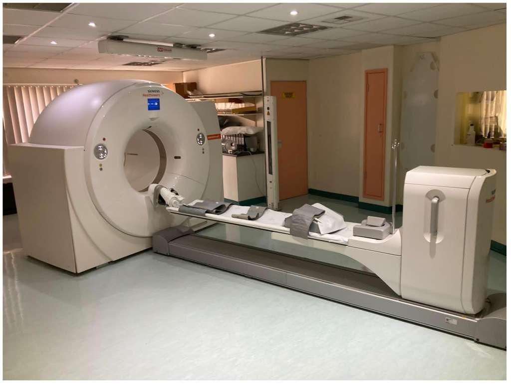
A modern PET CT scanner, which combines functional PET imaging with anatomical CT imaging in a single session
PET (The “Function” Scan)
Positron Emission Tomography (PET) is a type of nuclear scan that shows how your organs and tissues are working in real-time, not just what they look like.
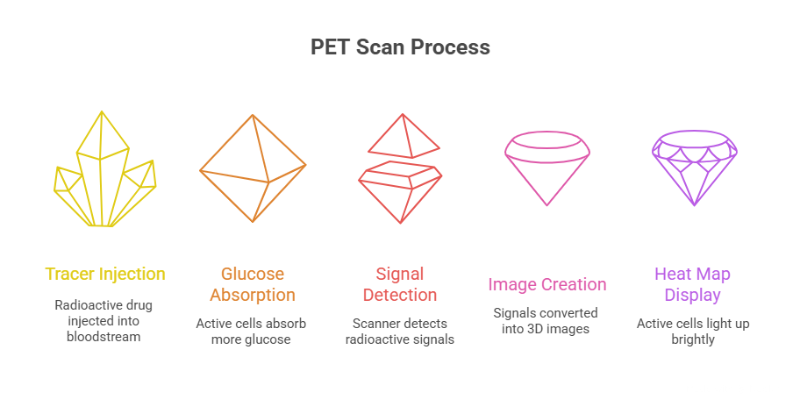
- The tracer: It is a special radioactive drug used in a PET scan. The most common one, called Fluorodeoxyglucose FDG, is basically a sugar mixed with a small amount of radioactive material. It’s injected into your bloodstream so the scan can track how your cells use energy.
- Here’s the principle: Your body’s cells need glucose (sugar) for energy. Cancer cells grow and divide faster than normal cells, so they use up more sugar. Because the tracer is made of glucose, these active cells absorb more of it and show up clearly on the PET scan.
- The “Heat Map” Analogy: Think of the PET scan as creating a heat map of your body. As the tracer breaks down, it releases tiny signals that the scanner detects and turns into 3D images. Areas that absorb more tracer—like active or abnormal cells—light up brightly, helping doctors spot changes often before other scans can Cleveland Clinic.
CT (The “Structure” Scan)
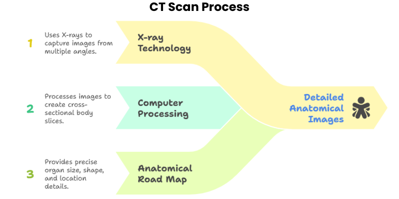
Computed Tomography (CT), sometimes called a CAT scan, uses special X-ray equipment to create detailed, cross-sectional images of the body’s internal structures. If PET shows function, CT shows anatomy.
- The Technology: A CT scanner takes a series of X-ray images from different angles around your body. A computer then processes these images to create cross-sectional “slices” of your bones, organs, and soft tissues.
- The “Anatomical Road Map” Analogy: A CT scan works like an anatomical road map of your body. It shows the exact size, shape, and location of your organs and can reveal problems like tumors with clear detail.
The Synergy (PET + CT): Fusing Function with Anatomy
The real power of a PET CT scan comes from combining both scans. The machine takes PET and CT images one after the other while you stay in the same position, then special software blends them into a single, detailed picture.
By merging the two scans into one image, doctors get both function and structure in a single view. This makes it easier to spot abnormal activity and pinpoint its exact location for a more accurate diagnosis Radiological Society of North America.
The PET scan can show a bright spot of unusual activity, but without CT, its location isn’t always clear. A CT scan can reveal a nodule, but not whether it’s cancer. Together, PET CT shows both — confirming abnormal activity, its exact location, size, and relation to nearby organs.
The PET CT Scan Procedure: A Step-by-Step Patient Walkthrough
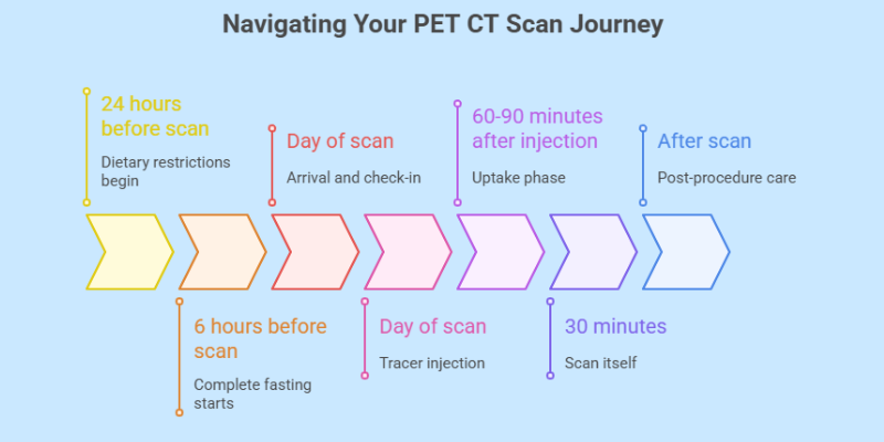
Understanding the process can significantly reduce anxiety. The entire appointment typically lasts between two to three hours, but the scan itself is much shorter. Here’s a chronological guide to what you can expect.
Before Your Scan: Preparation is Key
Proper preparation is not just a suggestion; it is essential for the accuracy of the PET CT scan. Failure to follow instructions can lead to a sub-optimal scan that may need to be repeated.
Communication with Your Medical Team
Before your appointment, it is crucial to inform your healthcare professional about several key factors. As advised by the Mayo Clinic, you should tell your team:
- If you have ever had a bad allergic reaction, especially to iodine-based contrast dye which is sometimes used for the CT portion.
- If you are pregnant, think you might be pregnant, or are currently breastfeeding.
- About any recent illnesses, infections, or medical conditions, particularly diabetes.
- A full list of any medications, vitamins, or herbal supplements you are taking.
- If you have claustrophobia (fear of enclosed spaces), as they may be able to provide a mild sedative.
Dietary and Activity Restrictions
The instructions you receive are designed to create the best possible conditions for the FDG tracer to work effectively.
- Diet: You will typically be asked to follow a very limited carbohydrate and no-sugar diet for 24 hours before your scan. For the final 6 hours before the exam, you must fast completely, drinking only plain water.
Why? As explained by cancer centers like CARTI, this is to lower your body’s normal blood sugar levels. If your blood sugar is high, your cells are already saturated with glucose, and they won’t absorb the FDG tracer as effectively. This can significantly decrease the quality of the images and hide abnormal cells. - Activity: Avoid strenuous exercise for 24 to 48 hours before the scan.
Why? Intense physical activity makes your muscles work hard and absorb a lot of glucose. If you exercise, your muscles will absorb the FDG tracer, which can create bright spots on the scan that could be mistaken for disease (a false positive) or obscure the view of actual problem areas. - Hydration: Drink plenty of water. Staying well-hydrated helps the tracer distribute evenly and makes it easier for the technician to place the IV line.
Special Instructions for Diabetics
Since a PET CT scan depends on how your body uses sugar, controlling blood sugar is very important for diabetic patients. Your levels must be within a safe range for the scan to work. The imaging center will give you clear instructions on food, medicine, and insulin timing—be sure to follow them closely and bring your diabetes medication to the appointment.
On the Day of the Scan: The Full Process
The PET scan process involves an IV injection of a radioactive tracer, a waiting period for absorption, and the scan itself
- Arrival and Check-in: When you arrive, you’ll check in and review the prep instructions with the technologist. You may need to change into a hospital gown and empty your bladder. A quick finger-prick test will also be done to check your blood sugar level.
- The Tracer Injection: For the tracer injection, a technologist will place a small IV in your arm or hand and inject the FDG tracer. You may feel a brief cool sensation, but the injection is quick and usually painless.
- The Uptake Phase (The Quiet Wait): After the injection, you’ll rest quietly in a dimly lit room for about 60–90 minutes. This waiting period allows the tracer to spread through your body and be absorbed by your cells. It’s important to stay still and avoid talking during this time.
- The Scan Itself: When it’s time for the scan, you’ll lie on a padded table that slides into the large, doughnut-shaped PET CT machine. The technologist will position you carefully, and it’s important to stay very still so the images come out clear. The scan takes about 30 minutes, is painless, and you may hear some soft buzzing or clicking sounds.
After the Scan: Post-Procedure Care
Once the scan is complete, the technologist will review the images to ensure they are clear. In most cases, you can leave immediately afterward.
- Flushing the Tracer: You will be strongly encouraged to drink plenty of fluids for the rest of the day. This helps to flush the remaining radioactive tracer from your body more quickly.
- Safety Precautions: The radiation from a PET CT scan is very low. As a precaution, you may be advised to avoid close contact with infants and pregnant women for about 6–8 hours after the test.
- Receiving Results: A radiologist will review your PET CT images and create a detailed report. This is sent to your doctor, who will schedule a follow-up appointment to explain the results and discuss your treatment plan.
Key Clinical Applications: When is a PET CT Scan Used?
PET CT scans are valuable because they can show how tissues are working, not just what they look like. They’re best known for their role in cancer care, but they’re also widely used in brain and heart conditions to help doctors diagnose and manage complex diseases.
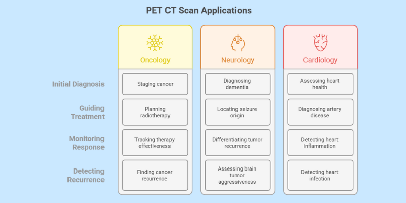
In Oncology (Cancer Care)
PET CT scans have transformed cancer care. They allow doctors to scan the whole body in one test, giving a clear picture of the disease PubMed Central. In cancer treatment, PET CT is used for:
- Initial Diagnosis and Staging: A PET CT scan helps doctors see if a lump or nodule is cancerous by checking its activity level. For example, a lung nodule that absorbs a lot of tracer often indicates cancer. The scan is also excellent for staging, showing whether the cancer has spread to lymph nodes or other organs—crucial information for deciding the best treatment.
- Guiding Treatment: PET CT scans are key in planning radiotherapy. They show the exact active tumor area, helping doctors target cancer more precisely while protecting healthy tissue. In fact, studies show PET CT can change treatment plans in about 1 in 4 patients by adjusting the dose or treatment area (PMC, 2010).
- Monitoring Treatment Response: During a course of chemotherapy or other systemic therapy, a follow-up PET CT scan can show whether a tumor is responding to treatment. A significant decrease in metabolic activity (FDG uptake) is an early sign of effective therapy, often appearing before the tumor physically shrinks on a standard CT scan.
- Detecting Recurrence: After treatment is completed, a PET CT scan is highly sensitive for detecting cancer recurrence, sometimes even before a patient develops symptoms. This allows for earlier intervention and potentially better outcomes.
Along with tracking treatment response, it’s also important to consider the emotional and mental health challenges that come after treatment. Read more about life after cancer and mental health here.”
In Neurology (Brain Disorders)
The brain is very active, it’s well-suited for PET imaging. Unlike MRI or CT, which mainly show structure, a brain PET scan shows how the brain and its tissues are actually working (MedlinePlus). Key uses include:
- Dementia and Alzheimer’s Disease: PET scans can spot reduced sugar use in certain brain areas, helping doctors diagnose different types of dementia early. With special tracers like Amyvid, they can also detect amyloid protein plaques—one of the main signs of Alzheimer’s disease (Alzheimer’s Society).
- Epilepsy: For people whose epilepsy doesn’t improve with medicine, surgery may be considered. A PET CT scan helps locate the exact area of the brain where seizures start, so surgeons can plan treatment that’s effective while protecting important brain functions.
- Brain Tumors: PET CT scans help doctors see how aggressive a brain tumor is. They can also tell the difference between a tumor that has come back and harmless changes from past radiation treatment, which often look the same on an MRI.
In Cardiology (Heart Conditions)
In cardiology, PET CT scans provide critical information about heart muscle health and blood flow that can guide life-saving interventions.
- Assessing Heart Muscle Health: After a heart attack, some heart tissue may be permanently damaged while other areas are still alive but not working properly due to poor blood flow. A cardiac PET scan can tell the difference (Weill Cornell HeartHealth), helping doctors decide if treatments like angioplasty or bypass surgery could restore heart function.
- Diagnosing Coronary Artery Disease (CAD): A cardiac PET scan shows blood flow in the heart at rest and during stress. It can clearly detect blocked arteries and is especially useful for patients where other tests don’t give clear results, such as those with higher BMI.
- Detecting Inflammation and Infection: PET CT scans are very effective at spotting heart muscle inflammation (like cardiac sarcoidosis) and infections linked to devices such as pacemakers or artificial valves—conditions that are often hard to diagnose with other tests (Mount Sinai).
Decoding the Cost of a PET CT Scan
For many patients, cost is a major concern. A PET CT scan is expensive because it requires advanced equipment, short-lived radioactive materials, and highly trained staff. The price can vary widely depending on several factors.
What Factors Influence the Price?
- Facility Type: This is often the biggest cost factor. PET CT scans done in hospitals are usually more expensive because of higher overhead and added facility fees, while outpatient imaging centers often charge less for the same test Capitol Imaging Services.
- Geographic Location: The price of a PET CT scan can vary by country, state, or even city. Scans in large metro areas usually cost more than in smaller towns or rural locations.
- Type of Scan & Tracer: A whole-body PET CT scan usually costs more than a scan of a single organ, like the brain. While FDG is the standard tracer, special tracers for conditions like prostate or neuroendocrine tumors are more costly and can raise the overall price.
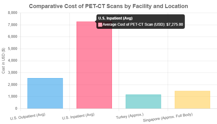
Note: Chart data is based on averages from sources including CareCredit, Istanbul Med Assist, and Health365 Singapore. International prices converted to USD for comparison. Actual costs will vary.
Actionable Advice: Before scheduling your scan, always contact your insurance provider to confirm coverage and request pre-authorization. Ask for a detailed estimate of your out-of-pocket expenses. If you are uninsured, you have the right to request a “Good Faith Estimate” from the healthcare facility before your procedure.
Weighing the Pros and Cons: A Balanced View of PET CT
Like any medical procedure, a PET CT scan has significant benefits but also comes with limitations and risks. A balanced understanding is essential for informed decision-making.
The Advantages (Pros)
- High Sensitivity & Early Detection: Its greatest strength is the ability to detect metabolic changes at the cellular level. This means it can often identify disease, particularly cancer, earlier than scans that rely only on detecting structural changes (Cleveland Clinic).
- Comprehensive Whole-Body View: In cancer care, a single PET CT scan can give a full-body picture of metabolic activity. This helps doctors see if cancer has spread, reducing the need for multiple separate scans.
- Enhanced Accuracy: By combining PET and CT data, doctors can pinpoint abnormalities more precisely. This leads to more accurate diagnoses, better treatment planning, and greater confidence in medical decisions.
- Painless and Non-Invasive: Aside from the brief discomfort of the IV injection, the scan itself is painless and non-invasive (Stanford Health Care).
The Limitations and Risks (Cons)
- Radiation Exposure: A PET CT scan does expose you to radiation from both the CT and the tracer. The dose is about the same as 8 years of natural background radiation American Cancer Society While not zero, the benefits of an accurate diagnosis usually far outweigh this small risk.
- Potential for False Results:
- False Positives: Not every bright spot on a PET scan means cancer. Infections or inflammation can also absorb the tracer and appear suspicious. That’s why doctors always interpret PET CT results along with your full medical history and other tests.
- False Negatives: A PET CT scan isn’t perfect. It can miss very small tumors or cancers that don’t absorb much tracer, such as some prostate, kidney, or mucinous cancers.
- False Positives: Not every bright spot on a PET scan means cancer. Infections or inflammation can also absorb the tracer and appear suspicious. That’s why doctors always interpret PET CT results along with your full medical history and other tests.
- Dependence on Blood Sugar: The accuracy of a PET CT scan depends on your blood sugar level. If you’ve eaten recently or have uncontrolled diabetes imaging centers, the results may not be reliable.
- Higher Cost: It is a more expensive imaging test compared to standalone CT, MRI, or ultrasound scans, which can be a significant factor for both patients and healthcare systems.
Future of PET CT: Compact and Smarter Scanners
PET CT technology is advancing rapidly. In August 2025, the U.S. FDA cleared a new compact multifunctional PET scanner developed by Brightonix Imaging. Unlike traditional large machines, this system is smaller, easier to install, and designed with patients in mind.
Key features include:
- Ability to scan different body regions (brain, chest, torso, extremities).
- Works for patients sitting or lying down.
- Compact design, making it accessible even for smaller hospitals and clinics.
- Built with advanced technology to improve precision and patient outcomes.
As demand for PET imaging continues to grow worldwide, such innovations may make PET CT scans more accessible, affordable, and widely available in the near future.
Frequently Asked Questions (FAQ) about PET CT Scans
Here are answers to some of the most common questions patients and their families have about the PET CT scan procedure.
Q1: Is the radiation from a PET CT scan dangerous?
A: The radiation dose is considered low and safe for medical diagnostic purposes. Medical professionals adhere to the principle of ALARA (As Low As Reasonably Achievable). The radioactive tracer has a very short half-life, meaning its radioactivity decays quickly, and it is naturally eliminated from your body within hours. Your medical team has determined that the benefit of getting an accurate diagnosis far outweighs the small associated risk. Young children and patients requiring multiple scans over their lifetime are treated with extra caution to minimize cumulative exposure (UT Southwestern Medical Center).
Q2: Is the PET CT scan painful?
A: The scan itself is completely painless. The only discomfort you might feel is a brief needle prick when the IV line is placed for the tracer injection, similar to a standard blood draw. The machine makes some noise, but it does not cause any physical pain.
Q3: What is the difference between a PET CT and a PET-MRI?
A: PET-MRI is a newer, emerging hybrid technology that combines PET with Magnetic Resonance Imaging (MRI). The main advantage of PET-MRI is that MRI provides superior soft-tissue contrast compared to CT, making it excellent for imaging certain areas like the brain, head and neck, liver, and pelvis. It also avoids the ionizing radiation from the CT scan. However, PET-MRI is currently less widely available, the scan takes longer to perform (around 45 minutes vs. 30 for PET CT), and it is often more expensive. PET CT remains the standard for most whole-body oncologic imaging (Cleveland Clinic).
Q4: Can a PET CT scan miss cancer?
A: Yes, it is possible. While highly sensitive for many common cancers, a PET CT scan can produce a false-negative result. This can happen if a tumor is too small to be detected or if it is a type of cancer with low metabolic activity (i.e., it doesn’t consume much glucose). This is why PET CT results are never interpreted in isolation. They are always correlated with your clinical history, physical exam, and the results of other diagnostic tests like biopsies and other imaging studies.
Q5: Will I be radioactive after the scan?
A: You will have a very small amount of radioactivity in your body for a few hours immediately following the scan. The tracer is designed to decay quickly. This is why you are advised to drink plenty of water to help flush it out of your system. The level of radioactivity is very low and poses no significant risk to you or those around you. The precautionary advice to limit close contact with pregnant women and small children for a few hours is simply an extra safety measure.
Conclusion: Making an Informed Decision About Your Health
The PET CT scan stands as a testament to the progress of medical technology, offering a unique and powerful window into the intricate workings of the human body. It is a cornerstone of modern medicine, providing critical information that shapes life-altering decisions.

Medical Disclaimer
This article is for educational purposes only and is not a substitute for professional medical advice, diagnosis, or treatment. Always consult a qualified healthcare provider before making medical decisions.