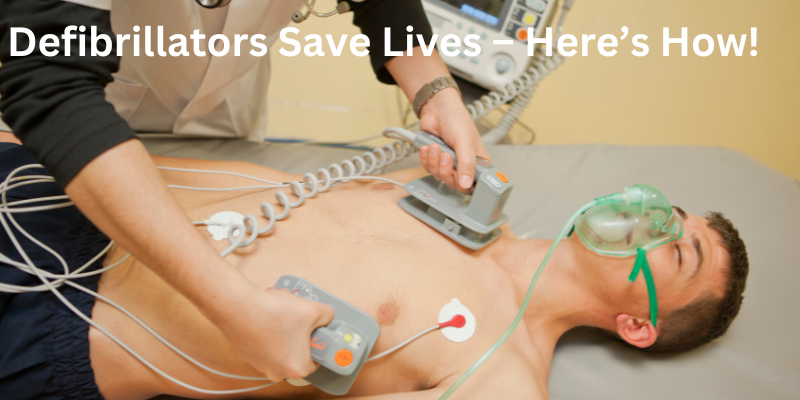
A defibrillator is a medical device that plays a critical role in treating life-threatening cardiac events, specifically ventricular fibrillation (VF) and pulseless ventricular tachycardia (VT). These conditions disrupt the normal rhythmic contractions(pumping) of the heart, leading to sudden cardiac arrest (SCA) and, if untreated it causes death. Defibrillators deliver a controlled electric shock to the heart, aiming to restore normal rhythm and save lives. This article delves into the clinical applications, underlying science, and technical aspects of defibrillators, along with insights into the cardiac conditions they address.
1. Clinical Use of Defibrillators
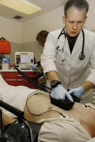
Defibrillators are employed in both pre-hospital and in-hospital settings to manage patients experiencing life-threatening arrhythmias. The devices are commonly seen in ambulances, emergency rooms, intensive care units, and even public spaces like airports and sports arenas. There are various types of defibrillators, each with specific applications:
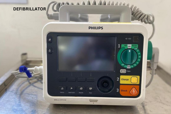
- Manual External Defibrillators (MEDs): Used by trained healthcare providers who can assess the patient’s condition and adjust the shock parameters accordingly.
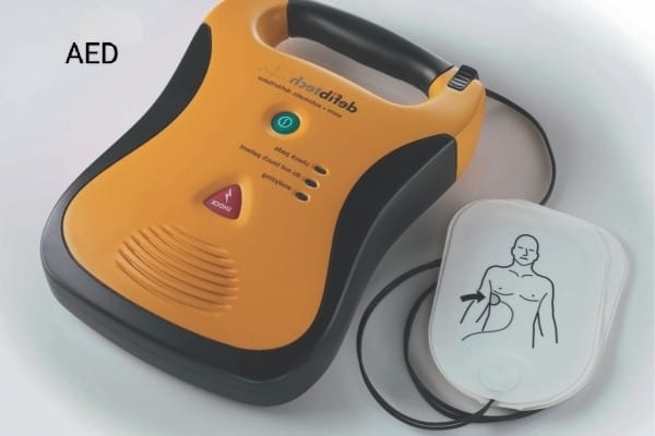
- Automated External Defibrillators (AEDs): Designed for use by the general public with minimal training. AEDs automatically assess the heart’s rhythm and determine whether a shock is necessary.
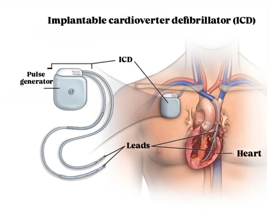
- Implantable Cardioverter Defibrillators (ICDs): Implanted in patients at high risk for cardiac arrest, ICDs monitor heart rhythm continuously and deliver shocks when necessary.
The primary goal of these defibrillators is to reverse fatal arrhythmias, such as ventricular fibrillation and pulseless ventricular tachycardia, before they result in irreversible damage.
2. Anatomy & Physiology of the Heart Related to Defibrillator Use
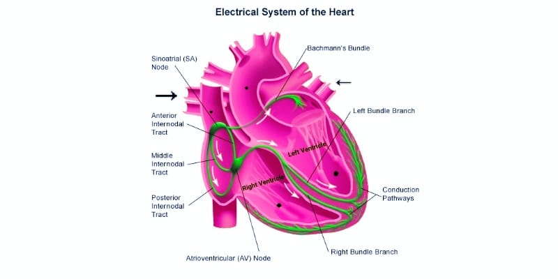
To comprehend why defibrillators are necessary, it is important to understand the Anatomy & Physiology of the Heart. The heart has its own electrical system, starting with the Sinoatrial (SA) node, which acts as the natural pacemaker and generates Electrical signals. These signals travel through the atria, ventricles, and conduction system. When the heart’s electrical signals become erratic, it leads to conditions like ventricular fibrillation (VF) or ventricular tachycardia (VT) which lead to sudden cardiac arrest (SCA) . A defibrillator shock resets this electrical activity, giving the heart a chance to beat normally again.
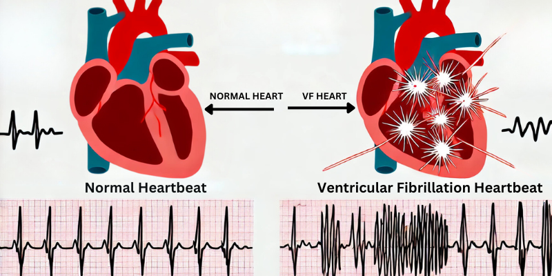
2.1 Ventricular Fibrillation (VF)
Ventricular fibrillation is a chaotic, uncoordinated contraction of the ventricles, leading to quivering rather than effective pumping of blood. VF is a medical emergency because it rapidly leads to a cessation of blood circulation, causing sudden cardiac arrest. During VF, the heart cannot generate a pulse, leading to loss of consciousness and eventual death if untreated.
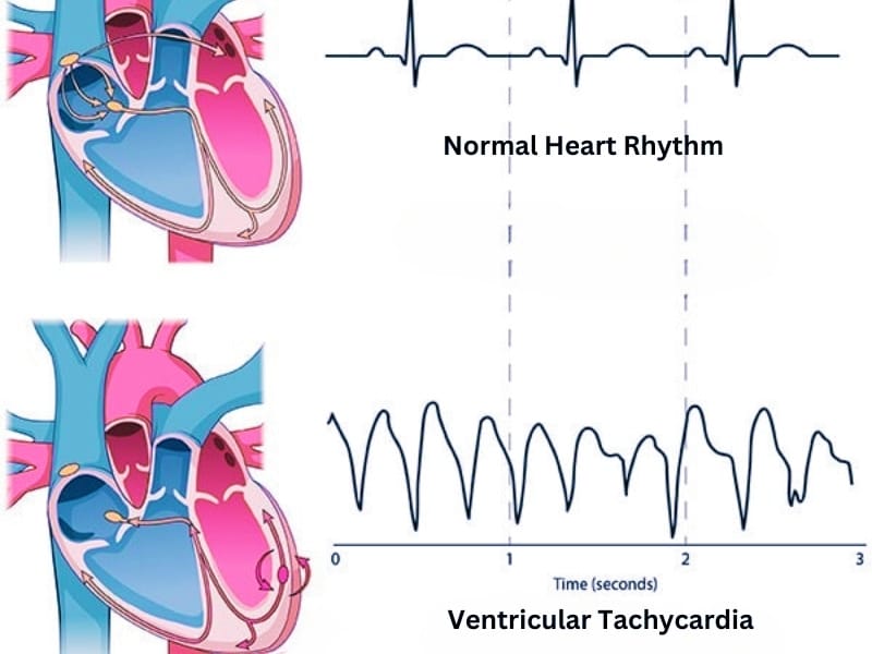
2.2 Ventricular Tachycardia (VT)
Ventricular tachycardia is a rapid heart rate that originates in the ventricles. It may cause a pulse, but in some cases, it progresses to pulseless VT, a life-threatening condition that can lead to VF. VT often arises due to scarring in the heart tissue, typically after a myocardial infarction (heart attack), disrupting the normal electrical conduction.
Both VF and pulseless VT require immediate intervention to prevent death, which is why defibrillation is critical in such emergencies
2.3 Sudden Cardiac Arrest (SCA): This occurs when the heart unexpectedly stops beating, often due to VF or VT, and requires immediate defibrillation and cardiopulmonary resuscitation (CPR).
3. The Science Behind Defibrillation
Defibrillators work by delivering a high-energy electric shock to the heart, aiming to depolarize the heart muscle cells in unison. This halts the chaotic electrical activity that characterizes VF and VT, allowing the heart’s natural pacemaker to resume normal rhythm.
3.1 Electrical Conductivity of the Heart
The heart is an electrically active organ, and its rhythm is controlled by the depolarization and repolarization of cardiac cells. Depolarization is the process by which cells become electrically activated, while repolarization restores their resting state. In VF or VT, the cells depolarize erratically, and the heart cannot pump blood effectively.
The defibrillator delivers a brief, high-energy shock that depolarizes all heart cells simultaneously. This action resets the electrical conduction system, allowing the sinoatrial node to regain control and restore organized contractions.
3.2 Shock Energy and Waveforms
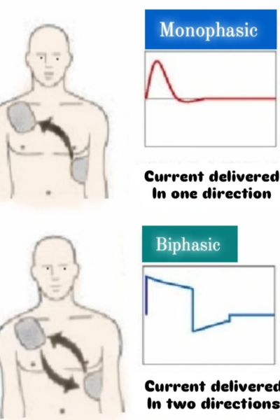
Defibrillation shocks are delivered using two types of waveforms:
- Monophasic Waveform: In earlier defibrillators, shocks were delivered in a single direction. This approach is effective but requires higher energy levels, which can cause more tissue damage.
- Biphasic Waveform: Modern defibrillators use biphasic waveforms, where the shock energy flows in one direction and then reverses. This technique requires less energy while being more effective at terminating arrhythmias, reducing the risk of heart damage.
Typical energy levels for defibrillation range from 150 to 360 joules, depending on the type of defibrillator and the patient’s condition.
4. The Importance of Defibrillation Timing
For all audiences, timing is the most critical factor in defibrillation. According to the American Heart Association, survival rates decrease by 7-10% for every minute defibrillation is delayed. This is why defibrillators, particularly AEDs, are placed in high-traffic public areas.
For medical professionals, the challenge is to respond swiftly with CPR and defibrillation to restore circulation before the patient suffers irreversible damage.
5. Advancements in Defibrillator Technology
Recent advancements in defibrillator technology have focused on improving efficacy, reducing side effects, and expanding accessibility. Innovations include:
- Improved Shock Algorithms: Advanced algorithms in AEDs and ICDs enhance the ability to accurately detect and treat arrhythmias while minimizing inappropriate shocks.
- Remote Monitoring: ICDs now offer remote monitoring capabilities, allowing physicians to track patients’ heart rhythms and adjust treatment plans accordingly.
- Miniaturization: The latest ICD models are smaller, more discreet, and easier to implant, improving patient comfort and reducing complication rates.
- Energy Optimization: Research continues into optimizing the energy levels used in defibrillation to balance effectiveness with reduced cardiac tissue damage.
6. Training and Education for All Stakeholders
To ensure defibrillators are used correctly and effectively, training is essential for all medical personnel, including doctors, nurses, and paramedics. They should regularly train on the use of manual defibrillators and integrate defibrillation protocols into broader emergency response efforts.
Biomedical engineers should regularly check devices, software updates, and pursue ongoing education on the latest technologies to ensure defibrillators remain in optimal working condition.
Conclusion :
- Sudden Cardiac Arrest(often due to VF or VT) is a common and unpredictable condition where the heart’s rhythm needs to be restored by an electric shock from a defibrillator.
- A defibrillator doesn’t restart a stopped heart. In fact quite the opposite, it actually stops a heart in the middle of a cardiac event, allowing the heart’s natural back-up system to take over and return it to normal sinus rhythm.
1 thought on “How Defibrillators Save Lives”