
Introduction: Peering into the Body’s Function
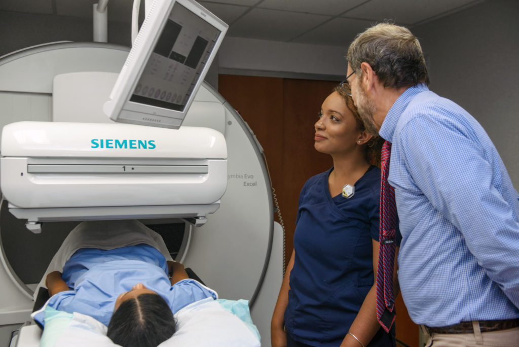
What if you could see not just the structure of an organ, but how it’s actually working in real-time? This isn’t science fiction; it’s the reality of nuclear medicine, and at its heart is a remarkable device: the gamma camera. Also known as a scintillation camera or, more famously, the Anger Camera, this technology has been a cornerstone of diagnostic imaging for decades, providing invaluable insights into physiological processes that other modalities can’t capture.
Invented by Hal Anger in the 1950s, the gamma camera doesn’t show you bones and tissues in anatomical detail like an X-ray or MRI. Instead, it creates images from the radiation emitted by a tiny, targeted radioactive tracer inside the body, revealing metabolic activity, blood flow, and cellular function. This article will demystify the gamma camera, exploring its core components, the elegant physics behind its operation, its critical clinical applications, and the exciting future of this essential nuclear medicine imaging equipment.
Table of Contents
How a Gamma Camera Works: The Anger Principle Explained
The genius of the Anger camera lies in its ability to convert invisible gamma rays into a visible image. The process, known as scintigraphy explained simply, involves several key steps and components working in perfect harmony.
- Radiotracer Administration: The process begins with the patient receiving a small, safe dose of a radioactive material called a radiopharmaceutical or radiotracer. This tracer is designed to accumulate in a specific organ or tissue. As it decays, it emits gamma rays.
- The Collimator: The Gatekeeper: Before the gamma rays reach the detector, they must pass through a collimator. Typically made of lead with thousands of precisely aligned holes, the collimator acts like a gatekeeper, only allowing rays traveling perpendicular to the detector to pass through. This filters out scattered radiation and is crucial for forming a clear image rather than a blurry mess.
- The Scintillation Crystal: Converting Energy to Light: After passing the collimator, the gamma rays strike a large, single crystal of thallium-activated sodium iodide (NaI(Tl)). This is the “scintillator.” When a gamma ray is absorbed by the crystal, its energy excites the crystal’s atoms, which then de-excite by releasing a flash of visible light—a process called scintillation. The brightness of the light flash is proportional to the energy of the gamma ray.
- Photomultiplier Tubes (PMTs): Detecting and Amplifying Light: Arranged in a hexagonal array behind the crystal is a set of 30 to 100 highly sensitive light detectors called photomultiplier tubes (PMTs). When the faint flash of light from the crystal hits the PMTs, each one converts the light photons into a small electrical signal.
- Positioning and Energy Logic: The “Brain”;: This is where Hal Anger’s ingenuity truly shines. The system’s electronics analyze the strength of the signal received by each PMT from a single scintillation event. The PMTs closer to the event produce a stronger signal. By comparing the outputs of all affected PMTs, the “Anger logic” circuitry can calculate the precise (X,Y) coordinate of where the gamma ray originally hit the crystal. The total sum of all PMT signals determines the energy of the event.
- Image Formation: Building the Picture: The system’s computer only accepts signals that fall within a specific energy window, corresponding to the radiotracer being used. This helps to reject lower-energy scattered photons. For every accepted event, a dot is placed at the calculated (X,Y) coordinate on a display. Over time (from seconds to minutes), hundreds of thousands of these dots build up to form a functional map, or scintigram, of the radiotracer’s distribution in the body.

Diagram illustrating how incident radiation creates light photons in a scintillator, which are then converted to an electrical signal and amplified by a photomultiplier tube (PMT)
Clinical Applications: Where Gamma Cameras Shine
The gamma camera is a versatile workhorse in hospitals worldwide, essential for diagnosing and managing a wide range of conditions. Its ability to visualize function makes it indispensable in many fields.
Nuclear Cardiology
This is one of the largest applications. Myocardial Perfusion Imaging (MPI) using Single-Photon Emission Computed Tomography (SPECT)—a technique where the gamma camera rotates around the patient to create 3D images—is used to assess blood flow to the heart muscle. It’s vital for diagnosing coronary artery disease, evaluating damage after a heart attack, and assessing heart function.
Oncology (Cancer Imaging)
Gamma cameras play a crucial role in oncology. Common uses include:
- Bone Scans: Highly sensitive for detecting if cancer has spread (metastasized) to the bones.
- Tumor Imaging: Certain radiotracers can identify specific types of tumors, such as in the thyroid or lungs.
- Sentinel Node Biopsy: Used to locate the first lymph node to which cancer cells are likely to spread from a primary tumor, guiding surgeons during removal.
General Nuclear Medicine
The applications are vast and varied, including:
- Endocrinology: Thyroid scans to evaluate for hyperthyroidism, hypothyroidism, or nodules.
- Nephrology: Renal scans to assess kidney function, drainage, and blood flow.
- Neurology: Brain scans to evaluate blood flow, which can be useful in diagnosing stroke, dementia, and other neurological disorders.
- Gastroenterology: Gastric emptying studies to measure how quickly food leaves the stomach.
The Evolution and Future of Gamma Cameras
While the fundamental Anger principle remains relevant, gamma camera technology is far from static. Continuous innovation is pushing the boundaries of what’s possible in nuclear medicine.
From Anger Cameras to Solid-State Detectors
The most significant recent advancement is the move from traditional NaI(Tl) crystals and PMTs to solid-state detectors, most notably Cadmium-Zinc-Telluride (CZT) detectors. CZT detectors convert gamma rays directly into an electrical signal, eliminating the need for PMTs. This results in higher sensitivity, superior energy and spatial resolution, and allows for much more compact camera designs.
Specialized and Portable Systems
The shift to CZT and other advanced technologies has enabled the development of highly specialized systems. Dedicated cardiac cameras, for example, are smaller, faster, and optimized for heart imaging, improving workflow in busy cardiology departments. Furthermore, new ultra-portable gamma cameras are emerging, bringing the power of nuclear imaging to the patient’s bedside, the operating room, or intensive care units.An example of a specialized system: the Digirad Cardius 2 XPO, a compact, dual-head gamma camera designed specifically for cardiac imaging
The Rise of Hybrid Imaging (SPECT/CT)
Modern systems frequently integrate a gamma camera with a Computed Tomography (CT) scanner. These SPECT/CT machines are incredibly powerful, as they overlay the functional data from the SPECT scan onto the detailed anatomical map from the CT scan. This fusion of function and form allows for precise localization of disease, dramatically improving diagnostic confidence.
Pros and Cons of Gamma Camera Imaging
Like any medical technology, gamma camera imaging has a unique set of strengths and weaknesses.
Advantages
- Functional Information: Provides unique insights into physiological processes, which is its primary advantage over anatomical imaging like CT or X-ray.
- High Sensitivity: Can detect molecular-level changes very early, sometimes before structural changes are visible.
- Versatility: Can be used to image nearly every organ system in the body with different radiotracers.
- Cost-Effectiveness: For many applications, it is a more affordable functional imaging option compared to PET.
Limitations
- Lower Spatial Resolution: The images are not as anatomically detailed as those from MRI or CT.
- Radiation Exposure: Involves a small dose of ionizing radiation, though it is generally considered safe and comparable to other imaging procedures.
- Scan Time: Imaging can take 30-60 minutes or longer, and the patient must remain still.
- Tracer Uptake Time: It can take several hours for the radiotracer to accumulate in the target organ before imaging can begin.
Frequently Asked Questions (FAQs)
How long does a gamma camera scan take?
The imaging itself typically lasts between 30 and 60 minutes. However, you must also account for the time it takes for the radiotracer to travel through your body and accumulate in the area of interest, which can range from minutes to several hours depending on the study.
Is a gamma camera scan safe?
Yes. While the procedure involves a small amount of radiation, the dose is carefully controlled and is considered safe. The benefit of an accurate diagnosis generally far outweighs the minimal risk associated with the radiation dose, which is often comparable to that of a standard CT scan.
What is the difference between a gamma camera and a CT scanner?
The fundamental difference is what they show. A gamma camera shows function—how an organ is working at a metabolic level. A CT scanner shows structure—the detailed anatomy and physical shape of an organ. This is why they are often combined in SPECT/CT systems to provide both types of information at once.
For a deeper comparison of functional imaging, you can also read our guide on PET CT Scan: Cost, Procedure, Meaning & Full Guide, which explains how PET/CT works and how it differs from gamma camera imaging.
What is SPECT?
SPECT stands for Single-Photon Emission Computed Tomography. It is an advanced 3D imaging technique that uses one or more gamma camera heads rotating around the patient. The computer then reconstructs the acquired 2D projection images into a 3D dataset, allowing doctors to view the organ’s function in cross-sectional slices, similar to a CT or MRI.
Conclusion: A Lasting Legacy of Innovation
From Hal Anger’s original design to today’;s advanced solid-state SPECT/CT systems, the gamma camera has remained a fundamental pillar of medical diagnostics. By allowing us to visualize the intricate functions of the human body, it provides critical information for diagnosing disease, guiding treatment, and monitoring patient health. As technology continues to evolve with advancements in detectors, software, and artificial intelligence, the gamma camera is poised to become even more powerful, precise, and integral to the future of medicine.
Have questions about gamma cameras or other medical technologies? As a biomedical engineer with deep expertise in this field, I’m passionate about sharing knowledge and fostering innovation.